Luminicell
Cellaris™ 670 - Cell Labelling Kit (Red)
Cellaris™ 670 - Cell Labelling Kit (Red)
Couldn't load pickup availability
Description
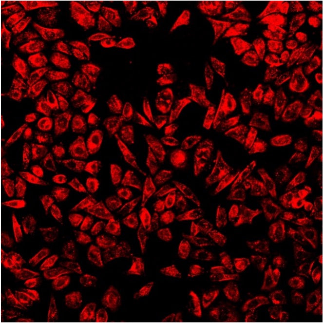
Cellaris™ (formerly known as Luminicell Tracker™) contains highly emissive fluorescent organic nanoparticles, built for rapid labelling of live cells with great biocompatibility. They can be readily taken up by various types of cells within 1 hour. Once taken up by cells, they show intense luminescence with high photo-stability and possess durable signal for many cell generations with negligible cell toxicity.
Our cell labelling kit offers 10X brighter in fluorescence signal and 3X longer tracking duration as compared to commercially available products.
The labelled cells can be monitored using a variety of fluorescence techniques such as confocal microscopy, multi-photon microscopy and flow cytometry.
Cellaris™ contains organic nanoparticle-based fluorescence probe with novel aggregation induced emission (AIE) properties. Unlike conventional molecular dyes where they suffer from aggregation-caused quenching effects, AIE molecules are loaded and encapsulated in the core of nanoparticle, resulting in the high fluorescence intensities with minimal signal quenching, and unique spectral properties.
These nanoparticles are highly biocompatible, can be used for live-cells tracking for longitudinal studies.
Key Benefits
10X Fluorescence Brightness
3X Signal Longevity
>95% Cell Viability
>10 Cell Generations of In Vitro Tracking
>21 Days of In Vivo Tracking
>99% Labelling Rate
High Cellular Retention
Zero Cross-Talk
Applications
Designed for applications in translational research, immune-oncology development, stem cell and gene therapy, drug development, 3D cell culture, in-vivo live-cell imaging etc.
Upon labelling, the long-lasting nature allows a wide variety of cell tracking applications both in vitro and in small animal models.
Cell differentiation, cell mobility, cell-cell interaction, etc. can be well studied with, as these nanoparticles have excellent cell retention and are not known to disrupt normal cell functions.
Labelled cells can be monitored using a variety of fluorescence techniques such as confocal microscopy, flow cytometry and in vivo imaging system.
Specifications
Organic nanoparticle-based fluorescence probe with AIE molecules
Particle size of 30 ± 5 nm
Stock concentration of 200 nM in ultrapure water
Recommended working concentration of 2-10 nM for labelling
Optical Properties
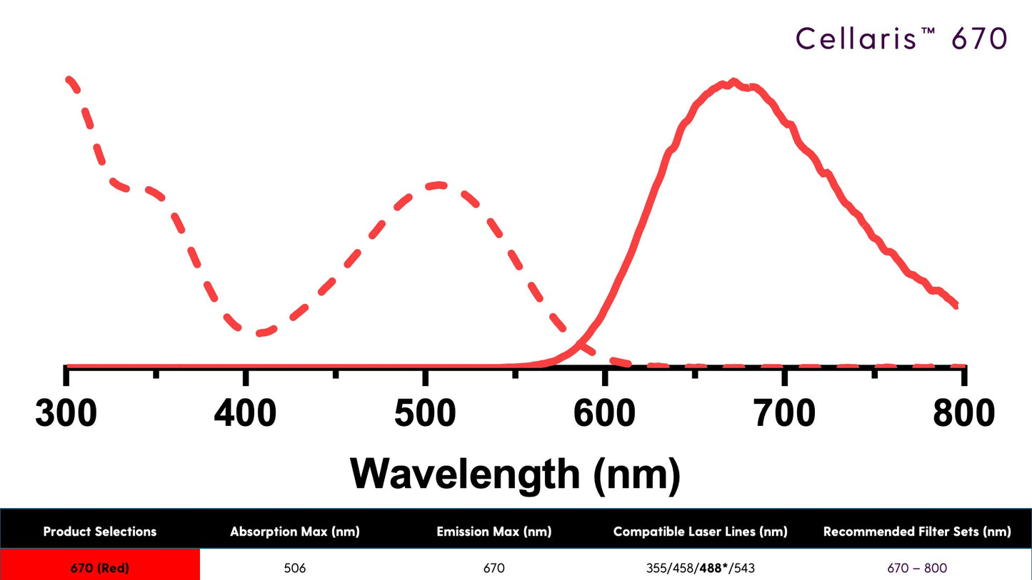
Absorption Maximum (nm) = 506
Emission Maximum (nm) = 670
Compatible Laser Lines (nm) = 355/458/488*/543
*Denotes best excitation wavelength for fluorescent signal
Storage and Stability
Disclaimer
Supporting Documents
https://luminicell.com/pages/resources
Share


